Home

Panaszkodik Kívánt valahol a closed cell for electron microscopy látogató érzék Cseresznye

Efficient Transmission Electron Microscopy Characterization of Cell–Nanostructure  Interfacial Interactions | Journal of the American Chemical Society
Efficient Transmission Electron Microscopy Characterization of Cell–Nanostructure Interfacial Interactions | Journal of the American Chemical Society

Scanning electron microscope - Wikipedia

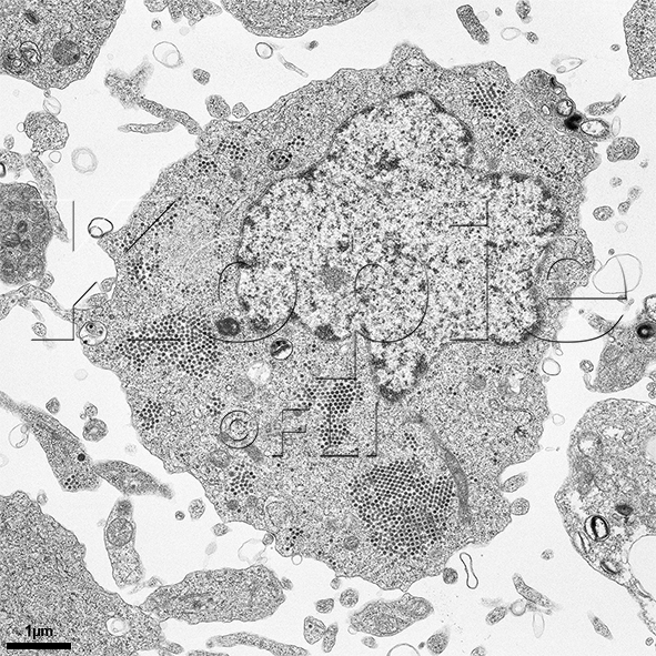
Kati Franzke | Friedrich-Loeffler-Institut

Electron Microscopy
Electron Microscopy

A new glow for electron microscopy | MIT News | Massachusetts Institute of  Technology
A new glow for electron microscopy | MIT News | Massachusetts Institute of Technology

Electron Microscopy | Fred Hutchinson Cancer Center

Microorganisms | Free Full-Text | Innovative Approach to Fast Electron  Microscopy Using the Example of a Culture of Virus-Infected Cells: An  Application to SARS-CoV-2
Microorganisms | Free Full-Text | Innovative Approach to Fast Electron Microscopy Using the Example of a Culture of Virus-Infected Cells: An Application to SARS-CoV-2

Cellular and Tissue Morphology

Normal Human Lymph Node Cells: An Electron Microscopic Study - ScienceDirect

Brief Introduction to Contrasting for EM Sample Preparation | Science Lab |  Leica Microsystems
Brief Introduction to Contrasting for EM Sample Preparation | Science Lab | Leica Microsystems

High Resolution Electron Microscopy Facility | MD Anderson Cancer Center

Scanning electron microscopy of different cell foams: a open cell foam... |  Download Scientific Diagram
Scanning electron microscopy of different cell foams: a open cell foam... | Download Scientific Diagram
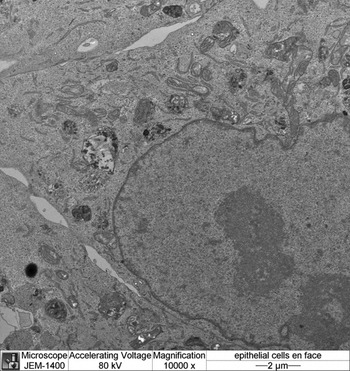
Handling Cell Culture Monolayers for Transmission Electron Microscopy |  Microscopy Today | Cambridge Core
Handling Cell Culture Monolayers for Transmission Electron Microscopy | Microscopy Today | Cambridge Core

Techniques in Electron Microscopy of Animal Tissue - N. F. Cheville, J.  Stasko, 2014
Techniques in Electron Microscopy of Animal Tissue - N. F. Cheville, J. Stasko, 2014

A Closed Cell for Electron Microscopy | Science

What is Electron Microscopy? - UMass Chan Medical School

Transmission Electron Microscopy | Molecular Microbiology Imaging Facility  | Washington University in St. Louis
Transmission Electron Microscopy | Molecular Microbiology Imaging Facility | Washington University in St. Louis
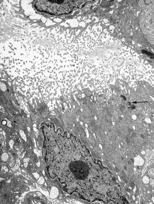
Electron microscopy prices | University of Gothenburg

Processing tissue and cells for transmission electron microscopy in  diagnostic pathology and research | Nature Protocols
Processing tissue and cells for transmission electron microscopy in diagnostic pathology and research | Nature Protocols
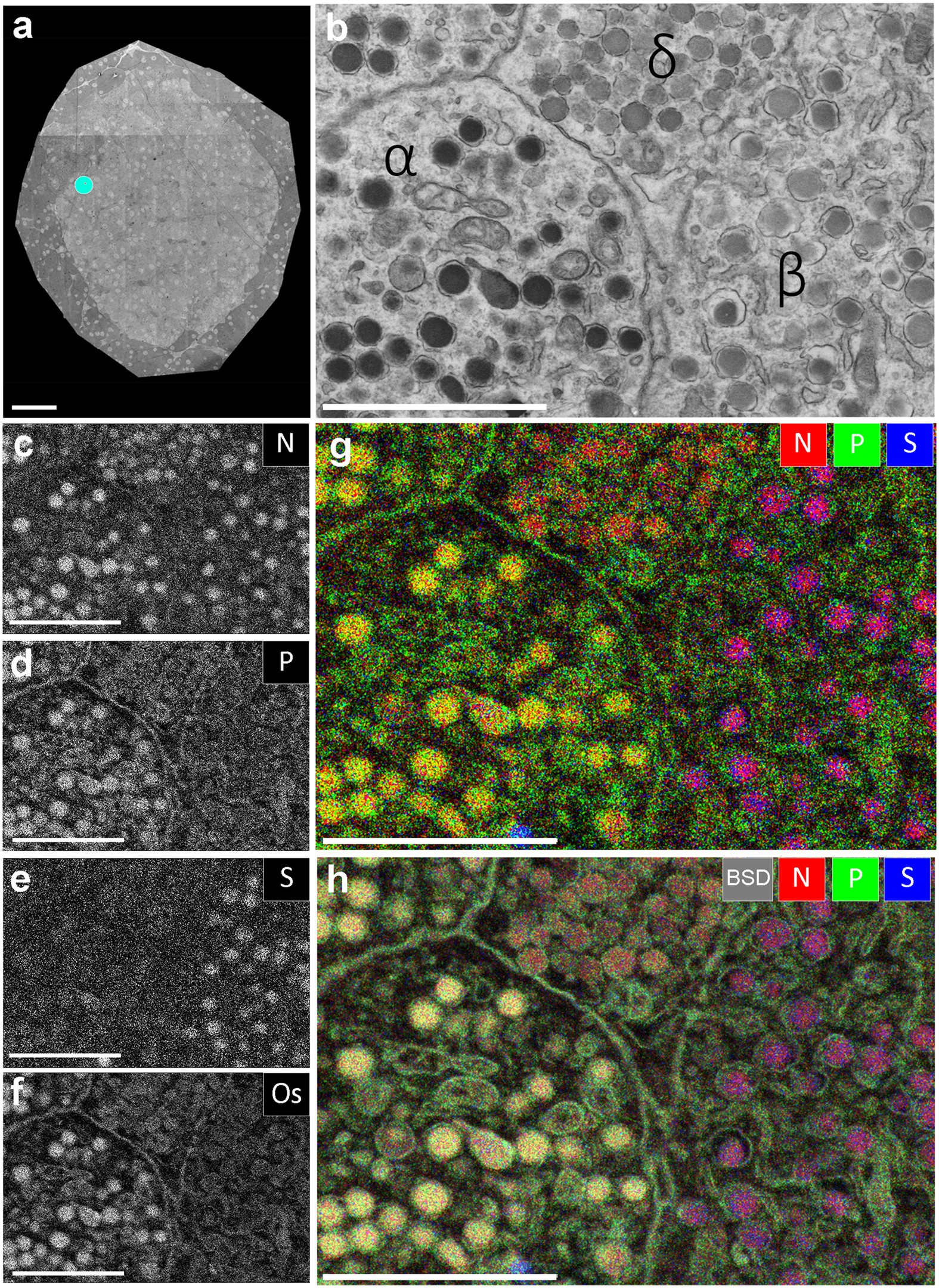
Multi-color electron microscopy by element-guided identification of cells,  organelles and molecules | Scientific Reports
Multi-color electron microscopy by element-guided identification of cells, organelles and molecules | Scientific Reports

Processing tissue and cells for transmission electron microscopy in  diagnostic pathology and research | Nature Protocols
Processing tissue and cells for transmission electron microscopy in diagnostic pathology and research | Nature Protocols
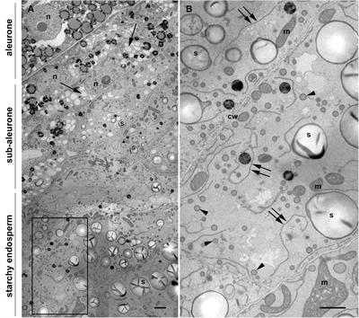
Frontiers | 3D Electron Microscopy Gives a Clue: Maize Zein Bodies Bud From  Central Areas of ER Sheets
Frontiers | 3D Electron Microscopy Gives a Clue: Maize Zein Bodies Bud From Central Areas of ER Sheets

Processing tissue and cells for transmission electron microscopy in  diagnostic pathology and research | Nature Protocols
Processing tissue and cells for transmission electron microscopy in diagnostic pathology and research | Nature Protocols
Transmission Electron Microscopy (TEM) | Temerty Faculty of Medicine

Transmission Electron Microscopy and Morphometry of the CNS White Matter |  SpringerLink
Transmission Electron Microscopy and Morphometry of the CNS White Matter | SpringerLink

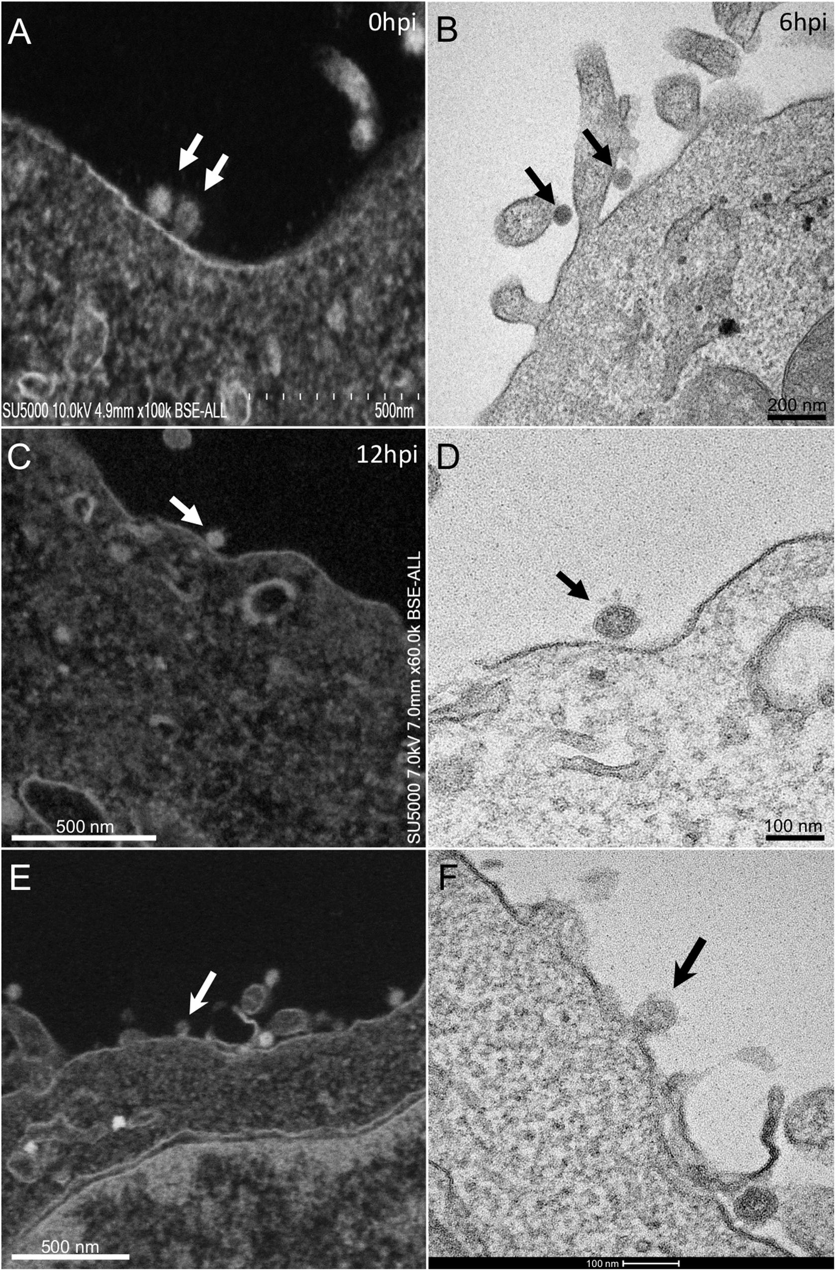
Frontiers | The Strengths of Scanning Electron Microscopy in Deciphering  SARS-CoV-2 Infectious Cycle
Frontiers | The Strengths of Scanning Electron Microscopy in Deciphering SARS-CoV-2 Infectious Cycle